
A mammogram, whether part of a routine screening or when used as a diagnostic exam, can help identify breast cancer. It can also identify other conditions, such as fibrocystic tissue.
A mammogram is a type of X-ray of the breast. Your doctor may order a screening mammogram as a routine check.
Routine screenings are an important way to establish a baseline of what’s normal. They can also be a tool for early diagnosis before you start showing symptoms of breast cancer.
Your doctor may also order a mammogram if have
After the test, a radiologist reviews the images and submits a report to your doctor.
The results are given a score of 0 to 6 under the Breast Imaging Reporting and Data System (BI-RADS). These categories help communicate the results and let the radiologist know what to look for in follow-up visits.
Read on to see example mammogram images and to learn more about normal and abnormal results look like.
Breasts contain fat, along with fibrous and glandular tissue. The more fibroglandular tissue you have, the denser your breasts are. The radiologist will classify your breast density using four categories:
- almost entirely fatty
- scattered areas of fibroglandular density
- heterogeneously dense
- extremely dense
When breasts are mostly fatty, the tissue on the mammogram is dark and transparent. That makes it easy to detect abnormalities, which generally show up as white.
Dense breast tissue appears solid white on a mammogram. Tumors and other masses also look white, so that makes it harder to detect abnormalities. Many women have dense breasts. Usually, but not always, your breasts get less dense as you age.
Some states require providers to notify women that they have dense breasts. If you receive such a notification, it doesn’t mean you have cancer or will develop cancer, although you’re at a slightly higher risk.
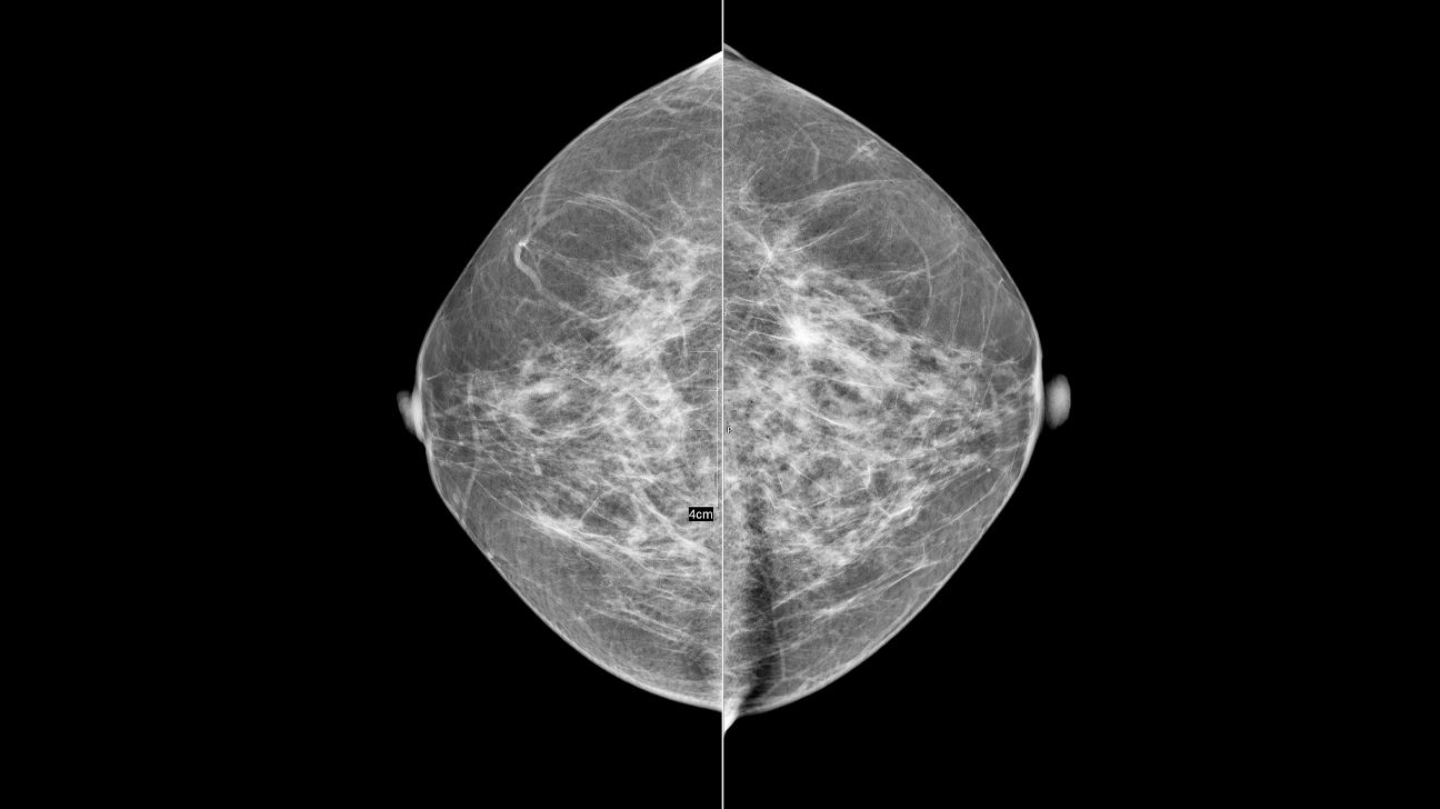
You can have dense breasts and still have a negative mammogram result. A negative result means nothing abnormal was detected. There were no distortions, calcifications, or lumps, and the breasts appear symmetrical. The BI-RADS score for this is 1.
Mammogram image of normal breast tissue
Deposits of calcium in the breast are called breast calcifications. They’re common on mammograms, particularly if you’re postmenopausal.
If you have calcifications, they’ll show up as white spots on the images.
Macrocalcifications look like large white dots or lines. They’re usually not cancerous. Microcalcifications look like tiny white specks, usually clustered together. Most are noncancerous, but sometimes they can be an early indicator of cancer.
This should be used for comparison each time you have a new mammogram.
You might also have a result of “probably benign,” with a BI-RADS score of 3. In fact, there’s a
Mammogram image of breast calcifications
Cancer isn’t the only reason you might have a lump in your breast. You can also develop fibrosis or cysts.
Fibrosis is when you have a lot of fibrous tissue. When you touch a fibrous area, it feels firm or rubbery.
Cysts are fluid-filled sacs that have a smooth, well-defined border. If a cyst grows large enough, it can stretch your breast tissue. When you feel a cyst, it’s usually soft, tender, and moveable.
Fibrocystic changes are more likely to occur during your childbearing years. They might be more noticeable just before you have a period. Like calcifications, fibrocystic tissue may have a BI-RADS score of 2 or 3.
Your doctor may want to order another mammogram or an ultrasound to investigate fibrocystic changes.
Mammogram image of fibrocystic breast tissue
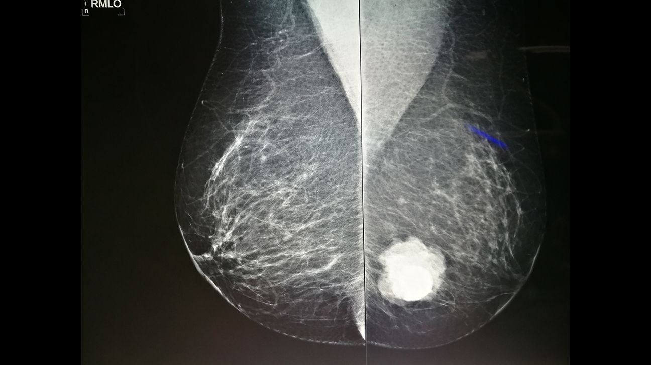
A cancerous breast tumor is usually irregularly shaped. Unlike a cyst, tumors are firm and they don’t move as freely. Most cancerous tumors are also painless.
If the radiologist sees a suspicious mass, they’ll give the mammogram a BI-RADS score of 4. That means it has an abnormality that doesn’t appear to be cancer, but that could be. They’ll probably ask you to have a biopsy to be sure.
When the image is highly suggestive of a cancerous tumor, the BI-RADS score is 5. That means the radiologist believes there’s a 95 percent chance that the tumor is cancerous. A biopsy is needed to confirm a diagnosis.
A BI-RADS score of 6 is only used when the tumor was already proven to be cancerous. Mammograms with this score are used to monitor breast cancer treatment.
Mammogram image of a breast tumor
If you have breast implants, you should still have screening mammograms. Even with implants, mammography is an effective way to screen for breast cancer. It’s harder to find abnormalities with implants, though. There’s also a slight risk of implant rupture during a mammogram.
You should mention your implants when making your mammogram appointment. Ask if the radiologist has experience performing and reading mammograms of women with implants.
Mention it again when you arrive for your mammogram. The healthcare professional may need to take a few extra images.
Mammogram image of breast implants
Talk to your doctor about your BI-RADS score. They can walk you through your results and make recommendations for future tests or treatment.
| BI-RADS score | What it means |
| 0 | The results are inconclusive or the radiologist wants another image for comparison. Radiologist will recommend another imaging study (mammogram or sonogram). |
| 1 | No abnormalities were detected. |
| 2 | Anything found, like cysts or calcifications, were benign. |
| 3 | Findings are probably benign. You may need follow-up imaging in 6 months. |
| 4 | An abnormality was found that could be cancerous but is most likely not. You may need a biopsy. |
| 5 | A tumor was identified with a 95 percent chance of being cancerous. You’ll need a biopsy. |
| 6 | A cancerous tumor has been confirmed. |
Mammograms are good at finding abnormalities before you can feel them. Early identification means treatment can begin sooner. Breast cancer is easier to treat before it spreads outside the breast.
However, mammograms can have false-negative results, meaning they miss some cancers. They may also produce false-positive results, which can lead to unnecessary biopsies or other procedures.
Overall, the accuracy rate is about 87 percent.
Getting called back after a mammogram doesn’t necessarily mean you have cancer. It does mean something needs clarification.
Sometimes, a mammogram will have a BI-RADS score of 0. It means you need additional imaging because the mammogram simply wasn’t clear enough to get a good reading.
A 0 score can also mean that the radiologist wants to look for changes by comparing older results to the current ones. This might be necessary if your previous mammograms were performed at a different facility and weren’t available to the radiologist. If that’s the case, you can request a record transfer.
Here are some of the reasons you might be called back:
- The images were of poor quality.
- The radiologist is waiting for prior mammogram results for comparison.
- The radiologist wants to get a closer look at breast calcifications, fibrocystic tissue, or other suspicious mass.
If cancer is suspected, your doctor will order additional testing, such as an ultrasound, MRI, or tissue biopsy.
Screening guidelines change as technology evolves and as we learn more about the benefits and risks of mammography. Currently, the
- Age 40–49 years: You should talk with your doctor to decide whether to start getting mammograms before age 50.
- Age 50–74 years: You should have mammograms every other year.
- 75 years and older: You should discontinue mammograms.
The
As long as you’re healthy and have a life expectancy of 10 years or more, you should continue breast cancer screening. Your doctor may recommend a different screening schedule or additional testing if you’re at a particularly high risk of breast cancer.
If you discover changes to your breasts, don’t wait for your next screening mammogram. Talk to your doctor right away.
What does cancer look like on a mammogram image?
Breast cancer on a mammogram can look like a white patch or mass. Cancers that begin in a milk duct don’t typically stay within the borders of the duct. Instead, they spread to nearby breast tissue and the outer edges look spiky or serrated rather than smooth.
What do cysts look like on a mammogram?
Large cysts can usually be seen on a mammogram. But smaller cysts, called “microcysts” may be difficult to detect. On a mammogram, large cysts appear as round or oval lumps with smooth edges.
Are white spots on a mammogram cancer?
Small, white spots on a mammogram that look like grains of salt are bits of calcium, called microcalcifications. They’re typically noncancerous, although certain patterns may be an early sign of cancer.
Your radiologist will have a better understanding of what they indicate, and may suggest additional scans if needed.
Does a shadow on a mammogram mean cancer?
Occasionally, you may be asked to return for additional scans if a radiologist notices a shadow on your mammogram images. Shadows don’t necessarily mean cancer.
Shadows can occur when areas of dense breast tissue appear together in one location. Shadows may also be due to a benign cyst. However, your doctor will likely ask for additional scans so that they can look at the area more closely and rule out the possibility of cancer.
If you have any questions at all about your mammogram results, talk to the doctor who ordered them. If your mammogram report references dense breasts, calcifications, or fibrocystic tissue, ask your doctor what that means and how it affects you.
Be sure to mention if you have symptoms or known risk factors for breast cancer, such as family history of the disease.